The Headwound As Seen At Bethesda Naval Hospital
For a more complete & detailed listing compiled by Dr. Gary Aguilar follow this link.
The photographs here have been stolen from Robert Groden's compendious photographic compilation in "The Killing of a President"
The purpose of this page is to illustrate the (apparent) incompatibility between the 'back of the head'
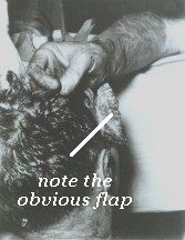
autopsy photographs (below) and the witness testimony. The autopsy photo shows the obvious and universally admitted
'right front' flap. The evidence is that there were in fact three
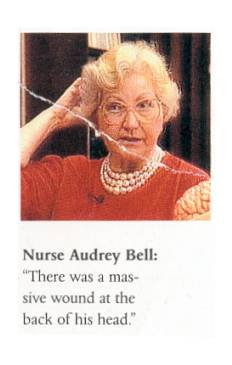
'major' flaps, one of them being at the right rear. (See Bell, Perry,
Grossman, McClelland, Peters below).
Dr Boswell - one of the autopsy doctors - maintains that he was lifting such a flap over the back of the head in the photo below.
 |
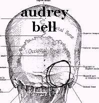
Drawing executed by Bell for the ARRB |
"Audrey Bell gave me a very explicit description of the flap, which was lifted for her
by one of the doctors to show her the large defect. She is very sure that it was hinged at the top, and not at
the bottom as in the photograph. She told me numerous times that "there was a lot of scalp missing in the
back though." Autopsy personnel have also described this flap in extensive detail. James Metzler described it in exactly the same place and facing the same way as did Bell, to both myself and Mark Crouch, in the studios of WCHE, Crouch's radio station, in West Chester, Pennsylvania, on May 1, 1991. The description was repeated to me in the same detail by Jerrol Custer.7 Dr. Robert McClelland also described this flap, but every one of these witnesses, and more, insists that it was hanging downward, hinged at the top to the left." (Harry Livingstone High Treason 2 p 316) "-Although only in Trauma Room One for 3-5 minutes, she did see the head wound. After asking Dr. Perry where is the wound, she said he turned the Presidents head slightly to the Presidents anatomical left, so that she could see a right rear posterior head wound, which she described as occipital in both her oral remarks, and in her drawings; -She said she could see brain and spinal fluid coming out of the wound, but could not tell what type of brain tissue it was; -She said it was her recollection that the right side of the Presidents head, and the top of his head, were intact, which is why she had to ask Dr. Perry where the wound was in the first place."(04/l 4/97 Summary of ARRB interview) |
| GENE AIKIN, MD: "The back of the right occipitalparietal portion of his head was shattered
with brain substance extruding." (WC-V6:65.) |
||
| CHARLES RUFUS BAXTER, MD: "...the right temporal and occipital bones were missing
(emphasis added) and the brain was lying on the table..." (WR:523). |
||
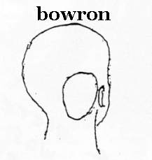 Drawing executed by Bowron. (Livingstone, 'Killing the Truth') |
NURSE DIANA HAMILTON BOWRON: "...there was blood all over this neck and shoulders. There
was a gaping wound in the back of his head." (Livingstone, Killing the Truth , p. 180) |
|
| KEMP CLARK, MD: Professor and Director of Neurological Surgery at Parkland, "...in the occipital region of the skull... Through the head wound, blood and brain were extruding... There was a large wound in the right occipitoparietal region, from which profuse bleeding was occurring... There was considerable loss of scalp and bone tissue. Both cerebral and cerebellar tissue were extruding from the wound." (WC--CE#392) |
||
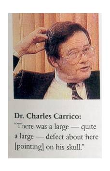 |
CHARLES JAMES CARRICO, MD: "The (skull) wound that I saw was a large gaping wound, located
in the right occipitoparietal area. I would estimate to be about 5 to 7 cm. in size, more or less circular,
with avulsions of the calvarium and scalp tissue. As I stated before, I believe there
was shredded macerated cerebral and cerebellar tissues both in the wounds and on the
fragments of the skull attached to the dura." (6H6) |
|
|
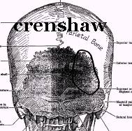
Drawing executed by Crenshaw for the ARRB |
CHARLES CRENSHAW, MD: It extended from the approximate center of the skull in the back to just behind the right
ear, utilizing a left to right orientation and from a position a couple of inches above the right ear to the approximate
middle of the right ear utilizing a top to bottom orientation." (FBI file # 89A-DL-60165-99) |
|
|
|
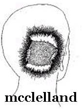
Don T. Curtis, D.D.S.: "The drawing by Dr. Robert McClelland is essentially my recollection of the wound suffered
by John F. Kennedy." |
|
| RICHARD BROOKS DULANEY, MD:" The wound was on the back of his head. On the back side"
They lifted up the head and "the whole back-side was gone." (Groden R., Livingston, H., High Treason.
1989 New York, Berkley Books, p.460.) |
||
|
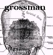
Drawing executed by Grossman for the ARRB |
ROBERT GROSSMAN, MD:"He (Grossman) said that he saw two large holes in the head, as
he told the (Boston) Globe, and he described a large hole squarely in the occiput,
far too large for a bullet entry wound...". " the second was a larger wound in the right parietal region (which he characterized as an exit wound) that was not an open hole in the cranium, but rather a plate of bone, about 6 cm in longest dimension, lifted up from the inside, which could really only be seen when Dr. Clark lifted up some of the Presidents hair" ( Grossman ARRB interview) |
|
| PAT HUTTON, RN : "Mr. Kennedy was bleeding profusely from a wound in the back of his
head, and was lying there unresponsive." (Price Exhibit V21 H 216). While helping with resuscitation
efforts a physician asked her to apply a pressure dressing to the head wound, she observed, however, that, "This
was no use, however, because of the massive opening in the back of the head."
(IBID) |
||
| MARION THOMAS JENKINS, MD: "a great laceration on the right side of the head (temporal and occipital) (sic),
causing a great defect in the skull plate so that there was herniation and laceration of great areas of the brain,
even to the extent that the cerebellum had protruded from the wound." (WC--Exhibit
#392) |
||
| RONALD COY JONES: ..".he had a large wound in the right posterior side of the head... There was large defect
in the back side of the head as the President lay on the cart with what appeared to be some brain hanging out of
this wound with multiple pieces of skull noted next with the brain and with a tremendous amount of clot and blood."
(WC-V6:53-54) |
||
 |
|
ROBERT McCLELLAND, MD: "...I could very closely examine the head wound, and I noted that the right posterior portion of the skull had been extremely blasted. It had been shattered...so that the parietal bone was protruded up through the scalp and seemed to be fractured almost along its right posterior half, as well as some of the occipital bone being fractured in its lateral half, and this sprung open the bones that I mentioned in such a way that you could actually look down into the skull cavity itself and see that probably a third or so, at least, of the brain tissue, posterior cerebral tissue and some of the cerebellar tissue had been blasted out...." (WC--V6:33) "There was no hole on that picture [the autopsy 'back of the head' pictures - see bottom of page ] that looked like that. And I said,. Well, I think I know what that is. I think it may be because if you notice there are some fingers at the top of the photograph apparently pulling a flap of scalp forward, and I think the flap was being pulled over that opening when they took the pictures." (ARRB) [McClelland surmises what Dr Boswell says was actually the case]
|
| MALCOLM PERRY, MD:: "I looked at the head wound briefly by leaning over the table and
noticed that the parietal occipital head wound was largely avulsive and there was visible
brain tissue in the macard and some cerebellum seen..." (HSCA-V7:302-interview with Purdy 1-11-78. "Writer Jimmy Breslin, who interviewed some of the Parkland doctors for an account published on Sunday Nov 24, reported the existence of a "huge flap" in the "occipito-parietal, which is a part of the back of the head." (Lifton, pp 321 pbk) An article appeared in some US papers on the 24th November 1963 as a "special from the New York Herald Tribune." It is obvious the author had interviewed Perry. In it, the author notes: QUOTE The wound in the throat was small and neat. Blood was running out of it, too fast. The occipito parietal, which is a part of the back of the head, had a huge flap." END QUOTE (Thanks to Barb Junkkarinen for this information.) |
||
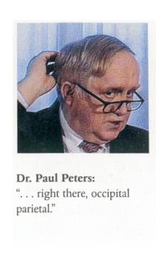 |
PAUL PETERS, MD:"...I noticed that there was a large
defect in the occiput...It seemed to me that in the right occipitalparietal area that there was a large defect."
(WC-V6:71) "The X rays of President Kennedy's skull, which we privileged to see later, showed dramatically how large the fragmentation of the skull was and was easily compatible with what Bob saw originally. There was a big hunk of bone sticking up there in the parietal.... I thought just exactly what Bob, did. They were probably making a series of pictures and they had just pulled that flap back up there to cover it up and took a picture of that to show the head with the flap restored, so to speak, for whatever reason. I'm sure there were many other pictures that were made at the same time. ...I think that [McClelland's drawing] pretty much corresponds to what I said, occipitoparietal. It looks a little further down in the occiput in this picture, I think, but it was pretty far posteriorly because you had to be able to see the cerebellum -- " (Peters, ARRB Parkland doctors interview) |
|
 |
||
|
" But the scalp was lacerated, & a pretty good sized piece of the frontal Q So you're saying that on the fourth view, which are the photographs that are in your hand right now, the scalp has been pulled back and folded back over the top of the head in a way different from the way that they appeared in the third view, the superior view of the head? A Yes. Q Is that fair? A In the previous one, it was permitted just to drop. In this one, it's pulled forward up over the forehead, toward the forehead. Q Who, if you recall, pulled up the scalp for the photograph to be taken?
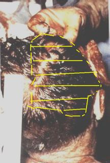
[Colour autopsy photo. The yellow hashed area marks the approximate location of the skull defect according to a skull Boswell marked for the ARRB ]
|
" Well, this was an attempt to illustrate the magnitude of the
wound again. And as you can see its 10 centimeters from right to left, 17 centi-
meters from posterior to anterior. This was a piece of 10 centimeter bone that
was fractured off of the skull and was attached to the under surface of the skull. . . There were fragments attached
to the skull or to the scalp and all the three
major flaps." (Boswell, interviewed by the HSCA FPP)
Boswell is explicit that the skull is missing beneath the scalp in the autopsy picture, above :
Q ...Now I'd like to ask you a question
about what is underneath the scalp of what we are
looking at now. Let's take the marking that
appears towards the hairline right at the base of
the neck, or where the hairline meets the neck. If
we take the point above that, where would you say
that the scalp is or that the skull will be missing
underneath the scalp that we can view there?
A Probably right about here.
Q So you're--
A Just about the base of the ear.
Q So you're pointing to approximately
halfway up the ruler that we can observe and to the
right of that small fragment, so the skull is
missing--
A Right.
(Boswell ARRB)
The Headwound As Seen At Bethesda Naval Hospital