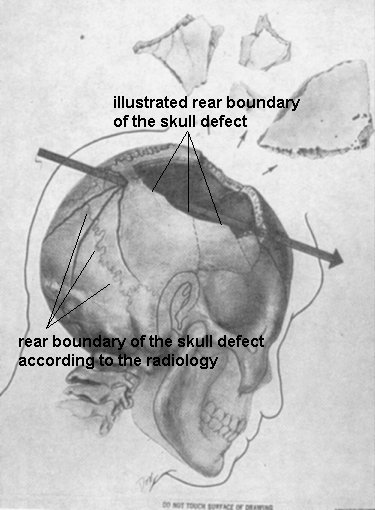
The Rear Boundary Of The Skull Defect
" With respect to the right frontoparietal region of the skull,
the traumatic damage is particularly severe with extensive
fragmentation of the bony structures from the midline of
the frontal bone anteriorly to the vicinity of the posterior
margin of the parietal bone behind "
(Clark Report)
" The findings and interpretation of the skull films are:
1. Nearly complete loss of right parietal bone..."
(G M McDonnel, Report for HSCA FPP)
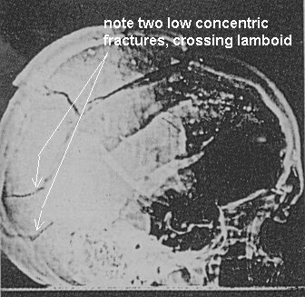
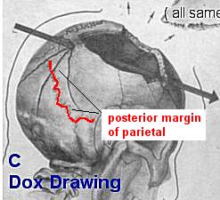
Here's the lateral x-ray . Note the two very obvious fractures indicated :
Now note that these fractures are conspicuously omitted from the Dox drawing. I have added their approximate locations
(in red) below :
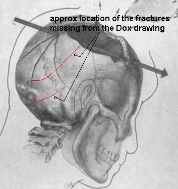
When we include these fractures, the rear skull looks like this (below) :
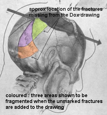
Note the low rear skull fragmentation pattern which results "to the vicinity of the posterior
margin of the parietal bone behind " as required by the Clark Report.
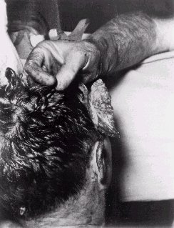
Below is one of the autopsy 'back of the head ' (BOH) photos showing the front right flap hanging open. Note it's size :
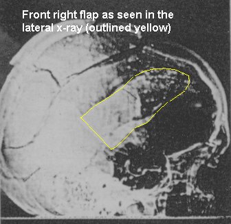
Here's the same flap, as seen on the lateral skull x-ray :
And here it is, seen in the Dox drawing :

It's shrunk.
A lot :-)
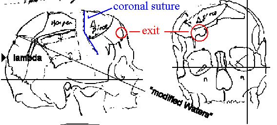
Below is the lateral x-ray, showing the location of the Clark Panel 'entry', just at the rear edge of a loose piece of bone sitting atop the skull :
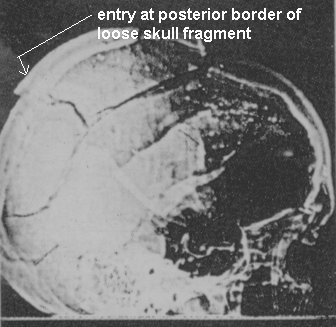
"On one of the lateral films of the skull (#2), a hole measuring approximately 8 mm. in diameter on the outer surface of the skull and as much as 20 mm. on the internal surface can be seen in profile approximately 100 mm. above the external occipital protuberance. The bone of the lower edge of the hole is depressed." (Clark)
So the entry (according to the Clark Panel) is at the rear end of this loose bone fragment.
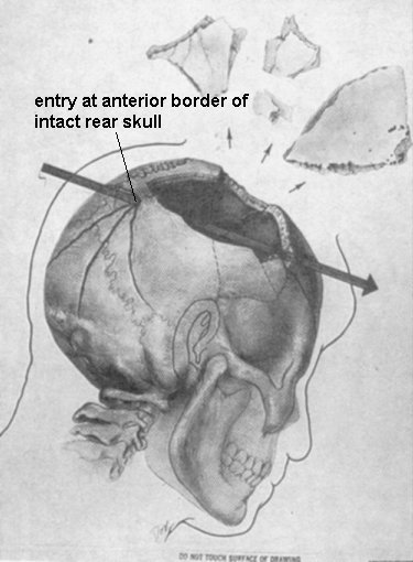
Below is the Dox drawing. Note that the entry is depicted in a perfectly intact (though fractured ) piece of bone. There is no sign of the lower edge of this entry hole being slightly anterior to any depressed fracture. In fact, there is no sign of any depressed fracture, period.
The Dox drawing and the Clark radiology cannot both be correct.
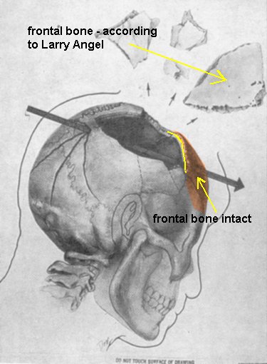
The Dox drawing shows the right frontal bone entirely intact. The coronal suture is marked yellow, below. The frontal bone is present & correct, just anterior to it. :
However, the Clark Panel opined that :
"With respect to the right frontoparietal region of the skull, the traumatic damage is particularly severe with extensive fragmentation of the bony structures from the midline of the frontal bone anteriorly to the vicinity of the posterior margin of the parietal bone behind " (Clark), so again, the Dox drawing and the ClarkPanel cannot both be correct.
Below is Larry Angel's drawing of the location of the loose bone fragments. The piece marked '[triangular'] piece'
is the large loose bone fragment I have indicated with the yellow arrow in the Dox drawing (above). Angel says
clearly that "Xrays 4, 5 & 6 show a large piece of skull vault , clearly frontal bone with
an apparent jagged line indicating coronal suture , about 7 to 8 cm long."
(Angel's report to the HSCA FPP)
Thus Angel has a large piece of frontal bone missing from the skull, whereas the Dox drawing shows no frontal bone
missing whatever.
The Dox drawing and Angel's report cannot both be correct.
Summary :
"The autopsy had been in progress for thirty minutes when 1 arrived. Cdr Humes
told me that he only had to prolong the lacerations of the scalp before removing
the brain. No sawing of the skull was necessary." ( Finck's Letter to Blumberg)
The picture below shows
A A recreation of the 'Dox skull defect' on a photo of the side of a human skull.
B A recreation of the defect, as shown by the radiology. ( The defect here is shown going back to the anonymous skull's lamboid.)
C The Dox drawing itself.
D The Dox drawing of JFK's brain.
All four images are to more or less the same scale.
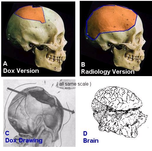
If we accept the 'Dox ' skull defect , the immediate question arises , how ( not to mention 'why' ) did Humes manage to remove the brain ( size shown bottom right) through a defect of the size shown in 'A' ?
The defect as determined from the radiology, being about 4 times the area, & extending from the front hairline to the very rear surface of the skull, does not lead to the same problem.
John McAdams has suggested that this page Brain Removal from some anatomy course material at the University of Vermont, may hold the answer to the 'brain removal' problem. It suggests, in relevant part :
"After reflecting the temporalis muscle tie a string around the skull about 1 inch above the imaginary line drawn between the inion, external auditory meatus, and the nasion. Mark this circumferential line with a pencil. This line indicates the proper level for cutting with the Stryker saw. A cut any higher than this presents a problem in trying to get your fingers under the temporal lobes of the brain."
The actual level of the cut recommended is shown below :
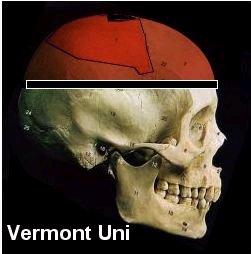
We now have the whole skull cap removed, as per the above image. This really is like the 'boiled egg with
the top sliced off" that SA Greer described seeing at the autopsy. Now compare what Vermont says is neccessary
to enable the brain to be removed to the defect shown in the Dox drawing :
The Dox version is nothing if not 'a cut higher than this' - in fact it involves less than half the 'skull removal'
the University of Vermont says 'presents problems'.
"Lift the temporal lobe of the brain medially so you can cut the tentorium cerebelli. Begin at the anterior
clinoid process and continue along the entire length of the ridge on the petrous portion of the temporal bone.
Continue until you come in contact with the occipital bone. " How on earth is Humes going to be able to reach
these structures through this hole ? :
He could hardly get *one* hand inside there (risking slicing his wrist on those jagged bone edges in the process
) let alone do anything constructive with it on the inside of the cranium.
"To remove the brain run your middle finger along the basilar artery down toward the spinal cord. Press down
with your middle finger as you retract your hand.
Make sure the tentorium cerebelli has been cut so that the cerebellum is free to come out."
The tentorium is located at a little below the level of the eop, deep inside the (supposedly) intact bowl of
the rear skull, right at the back..
The left rear brain ( occipital lobe) is seen to be absolutely intact in the brain drawings. ( see above)
To reach the tentorium, Humes must now somehow get the one hand he can get in there ( which fills the whole defect
so he can't even see what he's doing ) straight through the left rear brain ( without damging it in any
way ) to access the left side of the tentorium. In other words, he's cutting away blindly, with one hand, at something
he can't see, at something he can't get to anyway because the brain is in the way, while his wrist is slowly severed
by the jagged edges of bone around the defect.
Even if Humes still ( by some unaccountable miracle) managed to get the brain cut loose, he still would
not have been able to get the darn thing actually out of the cranium through that 4" hole.
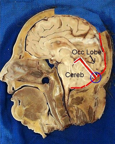
Picture above shows a sagittal section of human head. Part of the upper skull has been removed, as per the Dox drawing. ( 'Cowlick' to coronal suture) The occipital lobes were virtually intact according to Charles Petty (HSCA FPP). The blue circle shows where Humes must cut the attachment of the tentorium ( red & white line ) to the dura ( red line) in order to remove the brain.
Clearly. with the rear skull in this condition, there is no way to do this, as the occipital lobe(s) are between Humes & the point he has to cut.
Joe Riley (well known neuroanatomist) said some years ago what I
am pointing out here. If the boh was as 'intact' as the
Dox drawing might lead you to believe, it would be impossible to get the
brain out. (quote) :
" These interpretations fail to appreciate basic neuroanatomical
relationships (unfortunately, there was no neuroanatomist on either panel --
parietal foramina alone are enough to orient the photographs), are
contradictory, and ignore the obvious (it would be irresponsible
and stupid to try to remove the brain if so much skull were left, as it must
be in the official interpretations of the photographs) "
http://jfkhistory.com/riehl/What_Struck_John.html
( Riley holds a Ph.D. in Neuroscience, specializing in neuroanatomy and experimental neuropathology.
)
Paul Seaton
Jan 2004