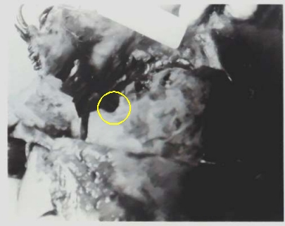
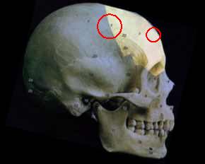
According to the FPP, there is a portion of a bevelled outshoot within the yellow circle (above).
Clearly, with an orientation of the photo, we can find the location of this formation. Dr Angel concluded as shown in his diagram below. (See his report). The 'exit' would then be within the red circle (below), inches forward of the coronal suture a few cm above the right eye socket, about the hairline.
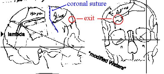
The FPP, for no stated reason, disregarded Angel, & put the 'exit' supposedly seen in the autopsy photo (above) way back in the coronal suture. (See below).
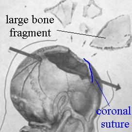
Note also the notation for a triangular 'piece' (the below fragment) on Angel's drawing, above.
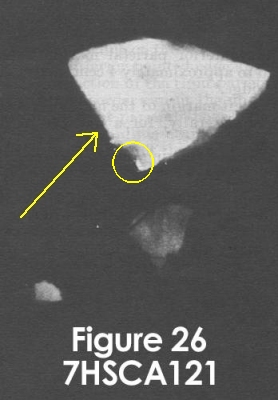
The above is the x-ray of the 'late arriving' bone fragments. At the circled corner of the largest of the three, the x-ray shows minute metal particles. Dr Angel found the large bone fragment to be 'clearly' frontal bone. He found that the circled area was located on the coronal suture. The image below shows the location of the frontal bone. Clearly, Angel puts the 'bevelling' seen on the autopsy photos nowhere near the 'exit site' suggested by the x-ray fragment. He has one in the hairline, and the other way back in the coronal suture (see the red circles marked below), with the large bone fragment - very unfortunately - falling between them. (See his diagram, above). Clearly if the autopsy photo 'exit' is any such thing, and if the 'exit' seen on the large fragment is ditto, and if Angel was right, then we are talking about two separate locations of exit. Since the headshot bullet fragmented, this would not be out of the question. However, the FPP does not appear to have been content with this situation, and went on the amalgamate the two 'exit' areas into one.
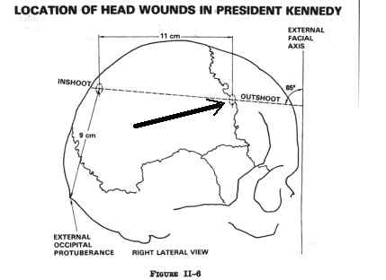
The drawing below shows the interpretation the FPP ended with. Note that the large bone frag (contra Angel) is now seen as coming from an area wholly behind the coronal suture - thus making a mockery of Angels' conclusion that the bone was 'clearly' frontal. By this sleight of hand, the 'offending' bone fragment is gotten out from between the two putative 'exit' locations. Angel's other location (in the hairline) is quietly moved back from the hairline to the coronal suture to amalgamate the two areas into one convenient but probably wholly inaccurate 'exit wound'.
Description:
In order to approximate the position of the two major loose fragments it is neccessary to define the gap seen in
xrays (esp. 1& 2 & photographs esp. 44 trans & photograph.) of the head & skull of JFK now kept
at the Nat Arch.
This gap where bone is missing along the top & right side of the skull vault extends
from just behind obelion( area of parietal foramina) forward almost to the frontal bosses anteriorly.
From the radio opaque lump behind the obelion which with cracks appears to mark te bullet entry the left margin
of the gap goes forward just to the right of the saggital suture
to a regio of major fracture just behind vertex where the margin moves about
1 cm to the left of the midline. From here the margin extends diagonally forward to the left to
a curved area about 5cm above the left orbit & about 5 cm from the midline.
The anterior edge of the gap crosses to the right , stepping down about the midline to a level 5cm above the nasion
and then sloping down to an area where there is almost semicircular
lacuna about 35 mm above the imddle of the right orbit.
To the right of this a vertical crack extends down to the orbit (an area of discoloration, apparently subcutaneous,
appears on the lateral photograph of JFK around the fronto malar angle of the right orbit.).
From a level about 4 cm above the frontomalar angle the bone margin extends backward on the right side , with another
v shaped crack in front of the coronal suture.
Behind this point the whole antero inferior quarter of the right parietal lies loose.
It's upper border was about 5cm above the squamous suture but in xray #? it appears shifted downward about 1cm.
From the point where it met the posterior half of the right parietal a big crack extends back & down , and
the posterior boundary of the gap goes backward & upward to the starting point just to the right of the obelion.
Large skull piece.
Xrays 4, 5 & 6 show a large piece of skull vault , clearly frontal bone with an apparent jagged line indicating
coronal suture , about 7 to 8 cm long.
The apparent inferior (right) border is 6cm long and at the irregular right angle which it makes with the jagged
(coronal) border are several radio opaque marks (part of a bullet?).
The third (anterior ) edge of the fragment is curvng. This large fragment
appears to be the upper part of the frontal bone, extending more on the right than the left, and leaving spaces
both in front & to the right.
The two smaller fragments in Xrays 4,5,6, are insufficient to fill these gaps.
Harper
The harper fragment photos show it as a roughly trapezoidal piece, 7 by 5.5 cm in size, coming mainly from the
upper middle third of the right parietal bone.
Near it's upper short edge vascular foramina on the inside & a faint irregular line on the outside indicate
saggital suture.
It's posterior inferior pointed angle appears to fit the crack in the posterior section of the right parietal and
it's slightlywavy lower border can fit the upper edge of the loose lower section of the right parietal.
It's upper short border , on the left of the midline near the vertex, may meet the left margin of the gap.
Behind it there appears to be a large gap, and in front a narrow one.